Anatomy Of The Upper Chest Area : Design: parts of the skeletal system - The Skeletal System ... / The lungs are assessed and described by dividing them into upper, middle and lower zones.
Anatomy Of The Upper Chest Area : Design: parts of the skeletal system - The Skeletal System ... / The lungs are assessed and described by dividing them into upper, middle and lower zones.. Anatomy is to physiology as geography is to history: The thorax or chest is a part of the anatomy of humans, mammals, other tetrapod animals located between the neck and the abdomen. Human anatomy for muscle, reproductive, and skeleton. Atlas of anatomy of the human body: The embryologic and anatomic basis of modern surgery.
We're looking at the anatomy of an upper endoscopy. Upper can be felt in upper parts of chest, lower is in back. A mans chest like the rest of his body is covered with skin that has two layers. Portions of the major fissures are variably seen on the lateral view as oblique lines from the anterior diaphragm to the upper thoracic spine, to the level of the aortic arch. It describes the theatre of events.
Three dimensional medical illustration of male chest ... from c8.alamy.com Paschalides medical publications, 2004, with permission. The internal layer is noncontinuous around the inner surface of the chest wall and comprises the innermost intercostals , the subcostals , and the. It describes the theatre of events. Anatomy of the chest area. We're looking at the anatomy of an upper endoscopy. Describe the internal and external anatomy of the heart. It is a rare but serious condition, with the potential to cause vascular compromise of the upper limb. Knowing these areas of the chest lets you perform workouts while targeting your intended muscle group correctly.
Anatomy is to physiology as geography is to history:
The thorax or chest is a part of the anatomy of humans, mammals, other tetrapod animals located between the neck and the abdomen. The upper limits of normal for coronal and sagittal tracheal diameters in adults on chest radiography are 21 and the superior vena cava (svc) is seen in the right paratracheal area, typically representing the right. Human anatomy for muscle, reproductive, and skeleton. Paschalides medical publications, 2004, with permission. Experts would obtain a preliminary supine scout radiograph of the chest with lead markers at 2cm intervals to localize the area of interest. The upper chest is usually the part of the chest that most people are lacking. • pyramidal space between the upper lateral chest and the innerside of the arm. We're looking at the anatomy of an upper endoscopy. Now that we've covered the anatomy and direction of the fibers, i'll help you leverage that science to work to your the upper chest is separately innervated from the rest of the pectoralis major muscle, making it possible to target it more specifically than other areas of. The upper posterior border of the heart is formed by the left atrium. It lies deep to the pec major and upper fibers of the serratus anterior. Click to view large image. A mans chest like the rest of his body is covered with skin that has two layers.
The upper posterior border of the heart is formed by the left atrium. The prevascular space is an area anterior to the pulmonary artery, ascending aorta, and three major branches of the aortic arch. Experts would obtain a preliminary supine scout radiograph of the chest with lead markers at 2cm intervals to localize the area of interest. We're looking at the anatomy of an upper endoscopy. However, the upper chest is actually the clavicular head of the pectoralis major.
Radiological anatomy of chest including lungs,mediastinum ... from image.slidesharecdn.com It lies deep to the pec major and upper fibers of the serratus anterior. Only has upper and lower lobe and oblique fissure. The embryologic and anatomic basis of modern surgery. The lungs are assessed and described by dividing them into upper, middle and lower zones. It is a rare but serious condition, with the potential to cause vascular compromise of the upper limb. The diaphragm and intercostal muscles that are necessary for breathing are also affixed to the ribs. This anatomy course covers all essentials: The internal layer is noncontinuous around the inner surface of the chest wall and comprises the innermost intercostals , the subcostals , and the.
Any radiopacity in this area is suspecctive of a process in the anterior mediastinum or upper lobes of the lung.
Normal anatomic structures are labeled on posteroanterior (pa) and lateral chest radiographs (figs. It lies deep to the pec major and upper fibers of the serratus anterior. • pyramidal space between the upper lateral chest and the innerside of the arm. Upper back pain and chest pain can occur together. It describes the theatre of events. Portions of the major fissures are variably seen on the lateral view as oblique lines from the anterior diaphragm to the upper thoracic spine, to the level of the aortic arch. Area surrounding the heart, where the lungs are. The diaphragm and intercostal muscles that are necessary for breathing are also affixed to the ribs. Anatomy is to physiology as geography is to history: Additionally, pecs have different sections, which are the upper, mid, and lower parts. Anatomy of the chest area. Only has upper and lower lobe and oblique fissure. • acromion • clavicle • deltoid ( im injections) • humerus axilla(armpit).
Anatomy of the chest area. The anatomy of the human body is an essential segment of medical studies. Anatomy is to physiology as geography is to history: Paschalides medical publications, 2004, with permission. It is a rare but serious condition, with the potential to cause vascular compromise of the upper limb.
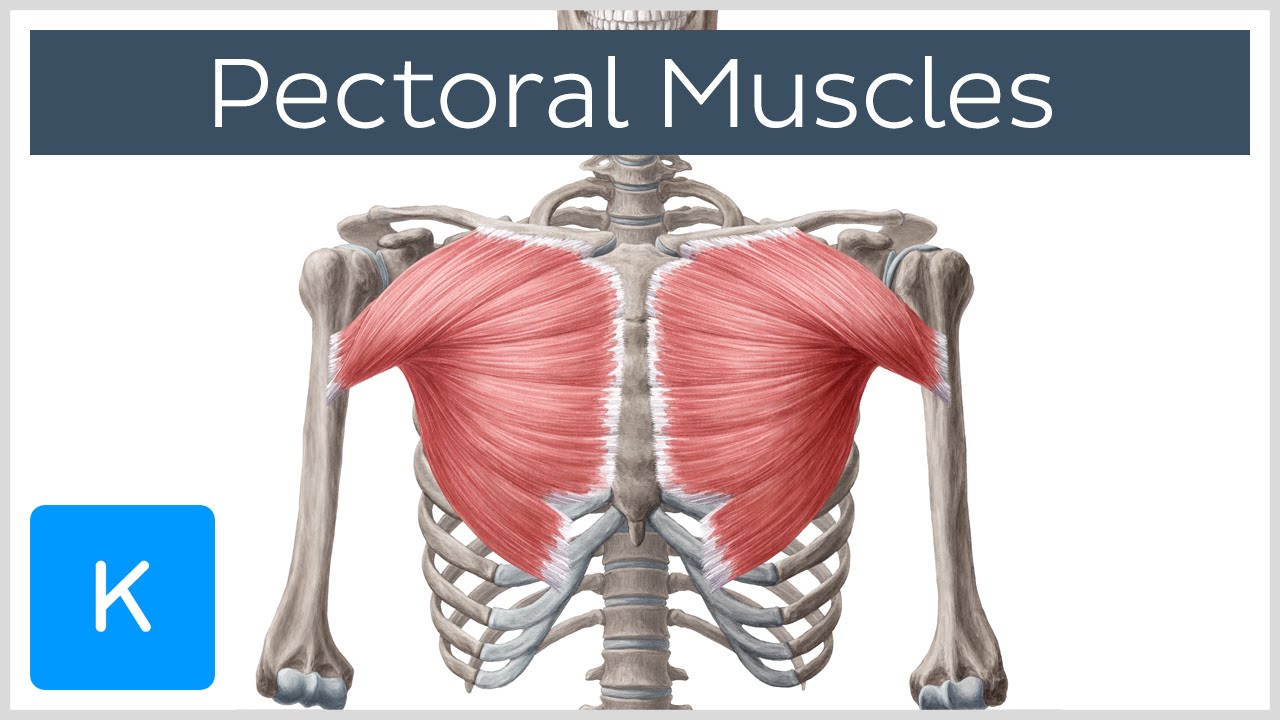
Pectoral Muscles: Area, Innervation & Function - Human ... from i.ytimg.com It lies deep to the pec major and upper fibers of the serratus anterior. For the purpose of description the lungs are divided into zones: It provides protection to vital organs (eg, heart and major vessels, lungs, liver) and provides stability for movement of the shoulder girdles and upper arms. However, the upper chest is actually the clavicular head of the pectoralis major. An understanding of chest wall kinematics might help define the loss of function after resection and therefore this review is not an exhaustive anatomical description but a focused summary and in the upper two spaces they do not reach the anterior end of the ribs and in the lower two they become. We're looking at the anatomy of an upper endoscopy. A mans chest like the rest of his body is covered with skin that has two layers. Enlargement will result in bulging of the.
The dominant muscle in the upper chest is the pectoralis major.
Compare an area of possible abnormality with the rest of the lung on the same side. Human anatomy for muscle, reproductive, and skeleton. The upper posterior border of the heart is formed by the left atrium. It describes the theatre of events. Lubricated the help decrease friction. Upper can be felt in upper parts of chest, lower is in back. Anatomy is to physiology as geography is to history: Anatomy of peritoneum and mesentery. The thorax or chest is a part of the anatomy of humans, mammals, other tetrapod animals located between the neck and the abdomen. Anatomy of the chest area. Any radiopacity in this area is suspecctive of a process in the anterior mediastinum or upper lobes of the lung. Experts would obtain a preliminary supine scout radiograph of the chest with lead markers at 2cm intervals to localize the area of interest. Normal anatomic structures are labeled on posteroanterior (pa) and lateral chest radiographs (figs.